Wrist Arthritis
Anatomy
Because of the number of bones in the wrist, the basic anatomy looks pretty daunting. Essentially though, there are two adjacent joints in the wrist, the radio-carpal and the mid-carpal joints (see figure).
Arthritis
The word “arthritis” literally means inflammation of a joint. This is a term that pre-dates x-rays and from a time when accurate diagnosis was not always possible. There are many different types of arthritis. The most common in the wrist is osteoarthritis, which is where the cartilage bearing surfaces of the joint have worn through. Often this becomes painful, once there is bone rubbing on bone.
While there are many causes of wrist arthritis, the most common cause is previous injury, either to a bone or a ligament. Both scaphoid malunions and scapho-lunate ligament injuries are a potent cause of wrist arthritis, and distal radius fractures affecting the joint surface can also lead to arthritis, sometimes even following optimal treatment.

Top: X-ray of wrist arthritis due to long standing scapho-lunate ligament deficiency – SLAC wrist, Bottom: X-ray following proximal row carpectomy – performed arthroscopically. The entire first row of the wrist bones have been removed, allowing contact between the healthy cartilage of the capitate and the radius.

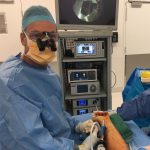
X-rays and photo of surgical wounds following an arthroscopic scaphoidectomy and mid-carpal fusion
Treatment
1. Wrist “simplification”
This involves surgery that will allow the wrist to move through one of the two joints, whilst removing the “bone on bone” joint. There are two forms of this approach – proximal row carpectomy and partial wrist fusions.
Proximal row carpectomy – in this operation, the first row of bones (scaphoid, lunate and triquetrum) are removed, leaving the capitate lying on the distal radius. For this operation to be effective, the cartilage on the capitate and the lunate fossa of the radius must be normal. It is also preferable that the end of the capitate has a round shape. In some people, the capitate has a flat or v shape, and is less suitable for this procedure. In my practice, an arthroscopic evaluation is important in assessing the cartilage and the suitability of the procedure. If all is good, the surgery may be performed arthroscopically in the same procedure. No metal-ware is required (see figure).
Partial wrist fusion – in these procedures, some but not all of the wrist bones are joined together with bony healing. There are various versions, depending on the pattern of arthritis which is present.
Scaphoidectomy with mid-carpal or “four corner” fusion. This is performed where there is arthritis between the scaphoid and the radius. The scaphoid is removed, to get rid of the “bone on bone” problem and to prevent the wrist collapsing, the mid-carpal joint is fused. This leaves the lunate free to move on the radius. Metalware is required. Again, suitability is best assessed arthroscopically (my preference at least) and the surgery can also be performed arthroscopically in the same procedure, with insertion of fixation screws through small incisions. Not all surgeons agree with this approach, as it is more time consuming than the open surgery and the longer term results are probably similar. However, idea appeals to me and some of my patients, and the early recovery does appear to be quicker with the arthroscopic approach (see figure).
Radio-scapho-lunate (RSL) fusion. This is used to treat arthritis between the radius and the scaphoid and lunate, usually when the lunate fossa is affected. Often this is after a fracture which has badly damaged the joint surface if the radius. Afterwards, movement will occur through the mid-carpal joint, so this must be in good shape prior to the fusion. The motion following the surgery can be improved by excising the end part of the scaphoid and even the triquetrum. However, later mid carpal arthritis may be associated with excision of the triquetrum. I perform this type of surgery as an open procedure, at this stage.
There are other forms of partial wrist fusion, such as mid-carpal (MC), scapho-capitate (SC), capito-hamate (CH), luno-triquetral (LT), scapholunate (SL), and scapho-trapezial-trapezoid (STT), but these are uncommon, and usually performed for non-arthritic conditions, such as instability or Kienbock’s disease.
2.Total wrist fusion
This involved making the wrist solid bone, from the radius to the metacarpal. Sometimes it is the only option. Although many patients don’t like the idea, which is understandable, when one has a stiff painful wrist, having a stiff painless wrist is not such a bad option. The surgery involves removing all the cartilage and subchondral (hard external) bone from the wrist bones, and compressing them together with a plate and screws, so that they heal together like a fracture. After the bones have healed, the plate is redundant, and can be removed if it is an irritation.
Total wrist fusion has been a reliable option for patients with wrist arthritis for many years.
3. Wrist denervation
This involves excision of nerves which carry pain from the wrist. A total wrist denervation requires a significant surgical exposure, I do not perform this technque. A partial wrist denervation is a much less invasive procedure, and can improve pain to a significant degree. In this procedure, two main nerves, the anterior and posterior interosseous nerves are divided through one or two small incisions. To predict the effect of this surgery, the nerves can be temporarily “blocked” with local anaesthetic. If the improvement in pain following the block is significant, then this suggests the surgery may be worthwhile. It is possible to try this surgery if arthroscopic assessment suggests that wrist simplification is not possible, and the patient does not want either a total wrist fusion or arthroplasty initially.
4. Arthroscopic debridement
“Cleaning-up” arthritic knee joints with arthroscopic surgery probably has no benefit. However, in the wrist, with certain conditions, such as early SLAC wrist (scapholunate advanced collapse), arthroscopic resection of 4mm of the tip of the radius may relieve bone on bone contact between the arthritic parts of the radius and scaphoid. A more aggressive “radial column resection arthroplasty” has also been described, with pleasing early recovery, and reasonable longer term results.
5. Wrist arthroplasty
Replacing the wrist joint with an artificial joint has had significant problems for many years. Typically, it has been rare to find a patient who is ideal for this procedure – an older lady who has painful osteoarthritis, with low demands, who doesn’t wish to loose motion with a fusion. More recently, a new design has become popular, with promises of improved longevity and durability. This has led to some surgeons using wrist replacements even in young, active male patients, with a technique that is relatively straight forward. Although the idea is appealing, I remain a little wary of this approach, especially when the wrist often lends itself to motion preserving partial fusions. Recent reports of failure of this type of joint replacement noted wear particles – both ketone and chromium, and suggested on going careful surveillance where these joints have been used, especially in young patients. Given the recent problem of chromium particles following some metal on metal hip replacements, this is not going to be my “go to” procedure for arthritic wrists. At least not for now.