Neonatal Brachial Plexus Injuries
Anatomy
The brachial plexus extends from the root of the neck into the upper arm. It is made up of nerves as they make their way from the neck into the arm and shoulder. Anatomically, it is complex, even in diagrammatic form, and in reality there is a high degree of variability in the fine details.
The brachial plexus transmits most of the muscle (motor) and sensory (feeling) between the spinal cord and the shoulder, hand, and arm.
Injuries to the brachial plexus vary according to the location and the severity, and this will determine the initial loss of function in the hand, arm and shoulder.
Birth injuries
Most brachial plexus problems in babies relate to injuries sustained at birth. Often the baby is heavier than average (more than 3.5kg). Frequently the baby gets stuck with their shoulder behind the mother’s pubic bone. This situation can be life-threatening to the baby, and consequently rapid relief of this problem is an emergency priority. Often this is when the nerves of the brachial plexus are stretched or more severely damaged. This sequence of problems is understandably very stressful for the parents.
Brachial plexus injuries can also occur during Caesarian section deliveries and even more rarely, by pressure on the plexus inside the uterus occurring prior to delivery.
Effects of injury
Initially, the obvious effect of the nerve injury is that some muscles do not function. Which muscles? Well, that depends on which part of the plexus is affected. Almost always, the shoulder muscles are affected, and typically the external rotators (which rotate the arm away from the midline) are weaker than the internal rotators. The elbow flexors are weak and sometimes the wrist extensors are also weak. This combination leads to the typical “waiter’s tip” position (see photo).
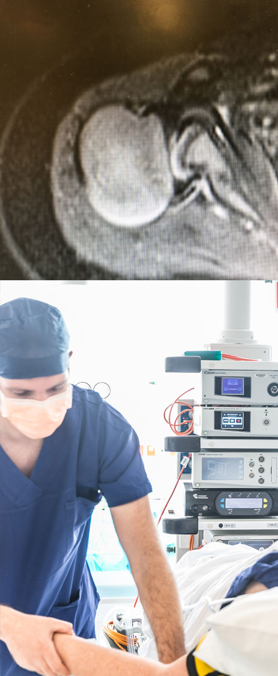
Top image: MRI of shoulder dysplasia following NBPI
The effects of the nerve injury can be very different from those in an adult. The violence of the injuries in babies is generally less severe than in those of adults, often caused by accidents like motorcycle crashes. So in babies, spontaneous recovery of nerve function is more likely. However, in babies growth is an additional factor. If a growing muscle is deprived of its nerve supply, it cannot lengthen normally afterwards. The longer the absence of nerve supply, the tighter it becomes later. This tightness combined with weakness of the muscle when it is paralyzed, can have profound effects on the shape of the shoulder joint, which can be permanent.
The most frequent persisting problems after NBPI occur around the shoulder. Generally the longer the nerve recovery takes, the more the shoulder is affected. However, significant shoulder problems can arise early on, and persist if untreated, despite the good recovery of nerve function.
Generally the elbow flexors recover spontaneously. In the past, great reliance was placed on the recovery of the elbow flexors, in trying to predict future function of the shoulder. This is largely because they share the same C5,6 nerve root inputs. If there was no recovery of elbow flexion by 3 months of age then it was thought that recovery of reasonable shoulder function was not possible without immediate nerve surgery. This has now be proven to be untrue. Early nerve surgery by no means guarantees good shoulder function, and good shoulder function is possible in many cases without early nerve surgery.
My preference is to focus on the shoulder from the outset. Whilst we do carefully monitor neurological recovery, we also careful monitor and treat the shoulder directly. There are a number of effective strategies to optimise the final outcome around the shoulder.
Evaluation
After finding out the history of the problem, most of the relevant information for the surgeon and therapists come from careful examination on a monthly basis. A pattern of recovery or lack of nerve recovery will be established, and the shoulder is carefully monitored for signs of tightness.
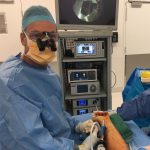
Some doctors use MRI of the plexus and or electromyography to help inform them of the severity of the problem. We have found that these tests are not more helpful than careful repeat examinations, and can also be misleading. The MRI scan requires a general anaesthetic in this age group, and in my opinion should be reserved for the shoulder, if it becomes tight and requires intervention.
It is impossible to reliably predict what treatment will be required or what the outcome will be on the first visit. Sometimes there is very rapid recovery, and it becomes pretty clear that the outcome will be excellent. Not uncommonly the recovery is gradual, with everyone anxiously waiting.
What we look for
Initially, we evaluate the level of injury in the plexus. We can tell this by which muscles are not working.
We also check for the following:
Horner’s syndrome: although rare, the eye can be affected, usually when the whole plexus is involved. This is often associated with a more severe injury, and if there is no return of function by 5 months, then early nerve surgery may be recommended.
Shoulder tightness: the shoulder can lose external rotation very early on. The therapists will show parents how to perform effective stretches to help prevent this, and monitor the passive motion every month.
If the shoulder becomes tight early on, for example at 6 months, then an MRI under general anaesthetic may be recommended. Generally, some form of surgical treatment may be required, either botox and casting, or sometimes surgical release, tendon transfers and casting. Additional nerve surgery may be considered, depending on the neurological recovery.
Muscle recovery: the therapists will perform muscle testing every month, using a special scoring system. A pattern of recovery can then be tracked.
Elbow flexion: it is encouraging to see return of active elbow flexion against gravity by 5 months of age. If there is no return of elbow flexion by 5 or 6 months of age, then nerve surgery may improve the final result for the child, however, this is quite uncommon.
Return of shoulder active external rotation: apart from maintaining supple passive external rotation, regaining active external rotation is very important in restoring good shoulder development and function.
If there is no return of active external rotation by 12 months of age, then nerve transfer surgery may be recommended to speed the recovery. This timeframe has been determined after careful analysis of the results of this surgery versus the natural history of spontaneous recovery.
If there is no return of external rotation by 24 months, and even if nerve surgery has been performed, then tendon transfer surgery may be recommended, to rebalance the joint, and aid in functional recovery.
Common surgical treatments
1. Botox and casting
Botulinum toxin is used to temporarily weaken the tight muscles, in kids where the internal rotators become tight, and are limiting passive external rotation. This is usually performed in children less than 12 months of age. The cast holds the shoulder in a position of external rotation. This may be left on for a number of months, especially if pre-operative imaging with MRI demonstrates significant changes in the shape of the shoulder socket (glenoid). We have found that these bony changes can occur in children as young as 6 months.
After application of the cast, a repeat MRI is usually performed to make sure the ball is well centered in the socket.
2. Nerve transfer surgery
In patients who fail to regain external rotation by 12 months of age, nerve transfer surgery can assist in the more rapid recovery of the main external rotator, infraspinatus. The lower part of the spinal accessory nerve is transferred into the suprascapular nerve, allowing motor axons to reinnervate the infraspinatus (see nerve injury section).
If there is absent elbow flexion at this time, then a nerve transfer may be performed for the elbow flexors.
Whilst excision of the injured part of the plexus and nerve grafting is still a reasonable option, it is my preference to attempt to bring healthy axons to the paralysed muscle as quickly as possible – hence nerve transfers. The sooner the muscle has a nerve supply, the better.
3. Rebalancing surgery
When the shoulder loses external rotation, or there is a failure of recovery of active external rotation, then rebalancing surgery may be the best option. This needs to be performed before there is an irreversible deformity of the shoulder joint, otherwise it will not work. Some older children who have normal shaped joints on MRI can be considered for this option, if they lack external rotation.
The surgery involves lengthening the tight internal rotator muscles and transferring the tendon of latissimus dorsi into infraspinatus. A cast is applied for 5-6 weeks afterwards. The result is fairly immediate, most families are happy with the outcome.
4. Humeral osteotomy
In older children who lack external rotation, and who have significant deformity of the shoulder joint, an osteotomy can really improve their function.
The patient must have sufficient internal rotation, as afterwards they will lose about 60 degrees of internal rotation. Essentially, the humerus is cut and the lower part externally rotated about 60 degrees. The two parts are then compressed and held with a metal plate and screws. Afterwards the arm is supported in a sling, and sports are avoided until there are signs of bone healing on X-ray. This usually takes about 8 weeks.